Anti-survivin antibody
Introduction:
Several proteins associated with malignant transformation of cells can induce auto-antibodies which are detectable in serum. Survivin, a novel member of inhibitor of apoptosis(IAP) protein family, suggested in both control of apoptosis and cell division. Survivin is not expressed in normal adult tissue expect for the thymus and placenta, but it is abundant in fetal tissue and various human tumors, including colon, lung, gastric, breast and bladder cancer. The enzyme-linked Immunosorbent assay(ELISA) is established for measuring anti-survivin auto-antibodies in human sera and evaluate the possibility of suffering from cancer. The kit with a low false positive rate is a good test for cancer screening.
|
|
 |
|
Principle:
| ELISA format |
|
 |
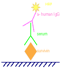
Survivin recombinant protein was coated on 96 well microplate for anti-survivin auto-antibody detection in human serum. The tracer is goat anti-human IgG F(ab')2 labeled with horseradish peroxidase. After adding tetramethylbenzidine (TMB) substrate and stopping by 1N HCl, the absorbent was measured at 450 nm for each well. |
|
|
Kit content:
1. Survivin ELISA plate: One strip holder containing 8x12 strips coated with survivin
2. Conjugate: one vial containing 11ml of HRP conjugated-goat anti-human-IgG antibodies.
3. 20x concentrated washing solution: one bottle, 20ml
4. Serum diluents: two bottles, 50ml each.
5. Positive control , 800 ul
6. Substrate (TMB): one bottle, 11ml.
7. Stop solution (1N HCl): one bottle, 11ml
8. One adhesive plate sealing film.
9. One sheet of semilog paper.
|
 |
|
ELISA procedures:
1. Take enough strips and put on strip holder.
2. Add 100 ul of serum diluents (blank), positive control sera and diluted serum samples to the individual well. Duplication is recommended for each control and blank.
3. Incubate at room temperature (21-25° C ) for 60 min.
4. Aspirate or shake out liquid from all wells. Add 280-300 uL of diluted 1X washing solution. Aspirate or shake out and turn plate upside down and blot on paper toweling to remove all liquid. Repeat the wash procedure 2 times (for a total of 3 washes).
5. Add 100 ul of conjugate to each well.
6. Incubate at room temperature (21-25° C ) for 60 min.
7. Wash with diluted 1X washing solution 3 times.
8. Add 100 ul of Substrate (TMB) substrate to each well.
9. Incubate at room temperature (21-25° C ) for 10 ± 2 min.
10. Add 100 ul of stop solution (1N HCl) to each well to stop the reaction. Tap the plate gently along the outsides, to mix contents of the wells.
11. Determine absorbance by reading assay plate at 450nm using 650nm as reference. The plate may be held up to 1 hour after addition of the Stop Solution before reading.
|
 |
Survivin antigen
Introduction:
Bladder cancer is a more common cancer now. At present, the best way to accurately diagnose bladder cancer remains performing cystoscopy and/or biopsy. It was demonstrated that survivin expression in bladder cancer (transitional-cell carcinoma, TCC) is an attractive prognostic marker for bladder cancer. The initial mechanistic studies found that survivin is cell cycle-regulated with a robust expression in G2/M phase.
|
| The localization of survivin which is associated with microtubules, centrosomes and mitotic spindles appears to be required for its function in regulation of apoptosis. Using survivin from urine as the basis of a potential bladder cancer screening test makes sense to us early on. All patients with bladder cancer had detectable survivin in their urine, while none of the healthy volunteers did. Easily degradation of survivin, the urine samples need to be centrifuged after collection and the sediment should be stored at -80℃. Lysis the cells of the urine sample before survivin protein before performing the enzyme-linked Immunosorbent assay (ELISA). Highly sensitive and specific determination of urine survivin appears to provide a simple, noninvasive diagnostic test to identify patients with new or recurrent bladder cancer. |
|
 |
|
Principle:
| ELISA format |
|
 |
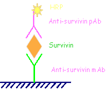
Cells are lysed for releasing survivin protein. Anti-survivin monoclonal antibody was coated on 96 well microplate for survivin protein detection in cells of urine samples. The tracer is rabbit anti-survivin polyclonal antibody labeled with horseradish peroxidase. After adding tetramethylbenzidine (TMB) substrate and stopping by 1N HCl, the absorbent was measured at 450 nm for each well. |
|
|
Kit content:
1. Survivin Microtiter Plate, One Plate of 96 Wells
3. Assay Buffer 20, 120 mL
4. Total Survivin Conjugate: anti survivin antibody-HRP, 11 mL,
5. Wash Buffer Concentrate, 100 mL,
6. human Survivin Standard, 500 pg/ tube
7. TMB Substrate, 12 mL,
8. Stop Solution 2, 11 mL,
9. Cell Lysis Buffer 2, 100 mL,
10. Protein assay reagent: coomassie blue
11. Protein concentration standard (BSA)
|
|
Storage:
All components of this kit are stable at 4 °C until the kit's expiration date.
|
 |
|
ELISA procedure:
1. Add 100 ul lysis buffer to each sample for releasing survivin protein in cells
2. Samples diluted 10X by sample diluent.
3. Take enough coated antibody strips and put on strip holder.
4. Pipet 100 μL of Standards #1 through #6 into the appropriate wells.
5. Pipet 100 μL of the Samples into the appropriate wells.
6. Incubate at room temperature (21-25° C ) for 60 min.
7. Aspirate or shake out liquid from all wells. Add 280-300 uL of diluted 1X washing solution. Aspirate or shake out and turn plate upside down and blot on paper toweling to remove all liquid. Repeat the wash procedure 2 times (for a total of 3 washes).
8. Add 100 ul of conjugate to each well.
9. Incubate at room temperature (21-25° C ) for 60 min.
10. Wash with diluted 1X washing solution 3 times.
11. Add 100 ul of Substrate (TMB) substrate to each well.
12. Incubate at room temperature (21-25° C ) for 10 ± 2 min.
13. Add 100 ul of stop solution (1N HCl) to each well to stop the reaction. Tap the plate gently along the outsides, to mix contents of the wells.
14. Determine absorbance by reading assay plate at 450nm using 650nm as reference. The plate may be held up to 1 hour after addition of the Stop Solution before reading.
|
 |
|
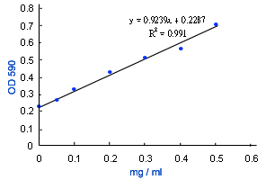
Total protein concentration for normalizing:
1. Cell lysate dilute 50X in PBS
2. Take non-coated strip and pipet 10 ul BSA standard diluted cell lysate in wells
3. Add coomassie blue at RT 10 min
4. Determine absorbance by reading assay plate at 590nm
5. Calculate the total protein concentration
|
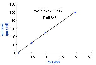
Survivin standard curve
 |
|
OD 450
|
pg/ml
|
|
0.068
|
0
|
|
0.512
|
25
|
|
0.964
|
50
|
|
1.975
|
100
|
|
|
Total protein standard curve
 |
|
OD 590
|
mg/ml
|
|
0.229
|
0
|
|
0.265
|
0.05
|
|
0.328
|
0.1
|
|
0.424
|
0.2
|
|
0.514
|
0.3
|
|
0.566
|
0.4
|
|
0.707
|
0.5
|
|
|
 |
Survivin Background
Introduction:
Survivin, a novel member of inhibitor of apoptosis(IAP) protein family, suggested in both control of apoptosis and cell division. The initial mechanistic studies found that survivin is cell cycle-regulated with a robust expression in G2/M phase.
The localization of survivin which is associated with microtubules, centrosomes and mitotic spindles appears to be required for its function in regulation of apoptosis. Survivin is not expressed in normal adult tissue expect for the thymus and placenta, but it is abundant in fetal tissue and various human tumors, including colon, lung, gastric, breast and bladder cancer. Several proteins associated with malignant transformation of cells can induce auto-antibodies which are detectable in serum. Overexpression of survivin by cancer cells may lead to anti-survivin antibody responses and cytotoxic T-lymphocyte responses against the cancer. Measuring anti-survivin auto-antibodies in human sera and evaluate the possibility of suffering from cancer. We will develop the ELISA kit for anti-survivin antibody detection, this kit with a low false positive rate is a good test for cancer screening.
Besides of the survivin auto-antibody detection, the survivin antigen detection could be applied as the specific tumor maker in specific sample. Such as detection of survivin in the urine sample assists in diagnosis of bladder cancer. Bladder cancer is a more common cancer now. At present, the best way to accurately diagnose bladder cancer remains performing cystoscopy and/or biopsy. It was demonstrated that survivin expression in bladder cancer (transitional-cell carcinoma, TCC) is an attractive prognostic marker for bladder cancer. Using survivin from urine as the basis of a potential bladder cancer screening test makes sense to us early on. All patients with bladder cancer had detectable survivin in their urine, while none of the healthy volunteers did. Highly sensitive and specific determination of urine survivin appears to provide a simple, noninvasive diagnostic test to identify patients with new or recurrent bladder cancer. |
|
 |
|
Principle (ELISA format kit) :
| Sera anti-survivin antibody detection |
Survivin antibody detection |
 |
 |
|
|